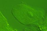
The buccal (cheek) epithelial cell is a standard specimen for light microscopy. These cells are readily obtained, by scraping the inner surface of one's cheek and putting the cells onto a glass microscope slide. The cells here are unstained, but viewed with differential interference contrast (DIC) optics. As before, the SD-9 is attached to the microscope via a T-mount adapter, which I fabricated, and a 1.4 x Sigma Teleconverter is installed between the T-mount adapter and the camera body. The thin depth of field allows for optical sectioning of the cell - this image is focused on the upper surface of the same cell, where ridges in the surface are readily visible. The companion image is focused so that the nucleus is clearly visible.
This image was only manipulated in Sigma Photo Pro, and adjusted for brightness and contrast. Sharpness was reduced in the program to -0.5.