


|
|
|
|
|
|
| Topics >> by >> all_about_lumbar_spinal_sten |
| all_about_lumbar_spinal_sten Photos Topic maintained by (see all topics) |
||
How The International Society for the Study of the Lumbar Spine can Save You Time, Stress, and Money.The posterior longitudinal ligament (PLL) affords only weak midline reinforcement, particularly at L4-5 and L5-S1, as it is a narrow structure connected to the annulus. The anterior and middle fibers of the annulus are most many anteriorly and laterally however deficient posteriorly, where the majority of the fibers are connected to the cartilage plate. 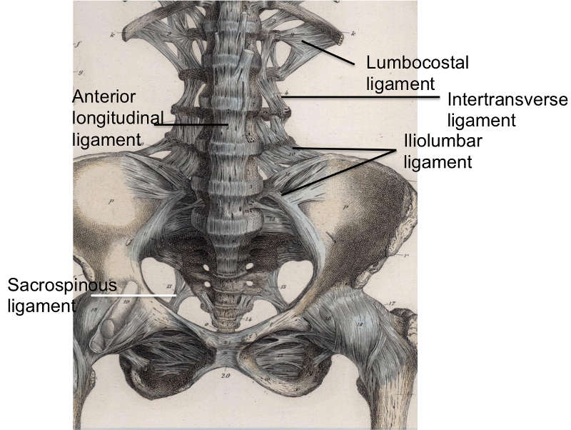 The anterior column (black dotted line) includes the anterior spinal ligament, the anterior annulus fibrosus (AF), the intervertebral disc, and the anterior 2 thirds of the vertebral bodies. The middle column (red dotted line) consists of the posterior aspect of the vertebral bodies, the posterior annulus fibrosus, and the posterior longitudinal ligament (PLL). ALL = anterior longitudinal ligament; ISL = interspinous ligament; LF = ligamentum flavum; NP = nucleus pulposus; SSL = supraspinous ligament. The annular fibers are strongly connected to the vertebral bodies and are organized in lamellae. This annular plan permits restricting vertebral movements, enhanced by investing ligaments. Lumbar Vertebral Ligaments The ALL covers the forward surfaces of lumbar vertebral bodies and discs.  Facts About Nomenclature and Classification of Lumbar Disc Pathology UncoveredThe ALL maintains the stability of the joints and limitations extension. The PLL is situated within the vertebral canal over the posterior surface area of the vertebral bodies and discs. It operates to restrict flexion of the vertebral column, except at the lower L-spine, where it is narrow and weak. Need More Info? joins the ideas of the spinous processes of surrounding vertebrae from L1-L3.  Often described together as the interspinous/supraspinous ligament complex, they weakly resist spine separation and flexion. The ligamentum flavum (LF) bridges the interlaminar period, attaching to the interspinous ligament medially and the aspect pill laterally, forming the posterior wall of the vertebral canal. It has a broad attachment to the undersurface of the exceptional lamina and inserts onto the leading edge of the inferior lamina. It maintains continuous disc tension. The intertransverse ligament signs up with the transverse processes of surrounding vertebrae and withstands lateral flexing of the trunk. The iliolumbar ligament occurs from the tip of the L5 transverse procedure and links to the posterior part of the inner lip of the iliac crest. It assists the lateral lumbosacral ligament and the ligaments pointed out above support the lumbosacral joint (see the following images). |
||
|
||