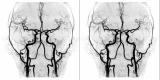
This image shows the blood vessels in the brain. The images have been made with an MRI scanner. To make these images it is not necessary to inject a contrast medium or dye. These images are called MIP's (Maximum Intensity Projections) and are part of a series where a projection is made every 10 degrees. Here two images are shown with a difference of 10 degree rotation. Technically speaking it is not a real stereo image, but when viewed with cross eyed technique one can see depth beautifully. This image is in video inverse signal, meaning the background is white , the blood vessels dark.