Vue partielle au microscope d'une préparation d'un ganglion lymphatique.
Grossissement : 50x.
Un colorant rouge a été employé pour permettre de voir la structure interne du ganglion.
La largeur de l'image ci-dessus représente environ 1,25 mm avec le dispositif électronique utilisé.
================================================================
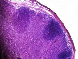
Partial view with a microscope of a lymph node.
Magnification : 50x.
A red coloring has been used here to see the internal structure of the lymph node.
The width of the above image represents about 1.25 mm with the electronic device used.